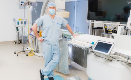
AI Predicts Best Test Option for Diagnosing Coronary Artery Disease

Thank you to the team at Hamilton Health Sciences for sharing this safety story with us. If your organization has a story, reach us at [email protected]. Through sharing and scaling lessons learned, together we can create the safest healthcare system.
Heart disease is the leading cause of death worldwide. The most common type is coronary artery disease (CAD), which occurs when the vessels that supply blood to the heart get narrowed, clogged or completely blocked by plaque – deposits of cholesterol, fats and other substances.
A test called an invasive coronary angiography is the current gold standard for diagnosing CAD. During this procedure, a catheter is inserted into the blood vessels through the patient’s groin or arm as a route to the heart. Then, a special dye that’s detected by X-ray is released in to the bloodstream. This allows doctors to see how the blood is flowing and identify any blockages.
“The problem is that our ability to determine which patients are best served by this test is poor and leads to a high rate of normal results, and potentially unnecessary procedures.”
Every medical test carries some level of risk. Globally, more than 50 per cent of patients who receive an angiography will have a normal test, showing their heart disease is not due to a blockage. This means these patients are exposed to unnecessary procedural risks. Plus, an angiography is a costly procedure that requires a highly skilled team.
To address this challenge, Dr. J.D. Schwalm, interventional cardiologist at Hamilton Health Sciences (HHS), and director of the HHS Centre for Evidence-Based Implementation, created and led a research trial.
Finding an Alternative
“About 90 per cent of patients referred for angiography have an appropriate referral based on clinical guidelines,” says Schwalm. “The problem is that our ability to determine which patients are best served by this test is poor and leads to a high rate of normal results, and potentially unnecessary procedures.”
The study looked at using a non-invasive imaging test called a coronary computed tomographic angiography (CCTA) instead of an angiography. When provided to the right people, the CCTA is a lower risk, highly accurate test that costs less. Radiation and X-ray dye-related risks are still present, but the procedural risks are eliminated. Study results showed that this approach could successfully reduce the number of angiographies by 76 per cent.
“The hardest part of the study was that we used an archaic system to pull out the right patients for CCTA. It was a labour-intensive process for the triage staff and cardiologists,” says Schwalm. “In practice, it’s not ideal.”
Improving the Process with AI
Bringing solutions to real-world health-care challenges is where collaboration between HHS’ CentRE for dAta science and digiTal hEalth (CREATE) and Centre for Evidence-Based Implementation come into play.
“This will help reduce the number of unnecessary angiograms, reduce health system costs and help ensure that patients receive the best test based on their risk for coronary artery disease.”
With funding from the trial, the team evaluated 10 years’ worth of patient data. Then, using artificial intelligence (AI) machine learning models, the CREATE team analyzed the data and determined the predictors of a normal angiography, thus determining which patients would and wouldn’t benefit from the procedure. The AI model provided a better prediction than existing clinical prediction scores and algorithms.
“We look forward to integrating the AI machine learning model into a decision support software tool that can be used by the referring physician or in the cardiac catheterization lab,” says Schwalm. “This will help reduce the number of unnecessary angiograms, reduce health system costs and help ensure that patients receive the best test based on their risk for coronary artery disease.”
Story by Ellie Stutsman, with photos by Josh Carey – Hamilton Health Sciences.
If your organization has a story, reach out to us at [email protected]. Together we can turn the corner on patient safety.
